







| Dental Podcast Episode - The Dr. Phil Klein Dental Podcast | Listen on: |

|

|

|

|
and others. |
Episode 801: Recognizing Risk: Oral Potentially Malignant Disorders Explained
 |
How confident are you that the white patch you noticed at your last exam wasn't something that needed a biopsy? The uncomfortable truth is that for every HPV-negative oral cancer, there was a visible precancerous lesion — and fewer than 5% of those lesions are identified before they become cancer.
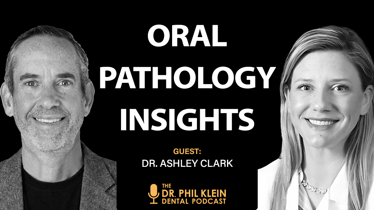
Dr. Ashley Clark is an Associate Professor and Division Chief of Oral Pathology at the University of Kentucky College of Dentistry. She holds a DDS from Indiana University and a certificate in Oral and Maxillofacial Pathology from the University of Florida. With over 40 published papers and abstracts, more than 200 continuing education courses delivered, a Fellowship in the American College of Dentists, a Fellowship in Health Education, and authorship of the oral pathology sections of both Dental Decks and Dental Hygiene Decks, Dr. Clark is one of the most prolific oral pathology educators in the country. She has earned multiple prestigious teaching awards, serves on the Commission on Dental Accreditation review board for oral and maxillofacial pathology programs, and sits on the Advisory Board for Oral Cancer Cause.
In this episode, Dr. Clark breaks down the full spectrum of oral potentially malignant disorders (PMDs) — from leukoplakia and erythroplakia to erosive oral lichen planus and proliferative verrucous leukoplakia — and explains exactly why the dental profession is failing to intervene early enough. The conversation covers the clinical criteria that demand a biopsy with no exceptions, why the standard two-week watch-and-wait approach is inappropriate for sharply demarcated lesions, and how to structure long-term surveillance once a PMD is diagnosed. Dr. Clark also addresses the medicolegal exposure dentists face when PMDs go unmonitored, and makes a clear, evidence-based case that appropriately treating leukoplakia alone could reduce oral cancer rates by 50%.
- A sharply demarcated white lesion requires a biopsy regardless of patient history, smoking status, or lesion duration — no additional clinical information changes this decision. Watching such a lesion without a diagnosis is not a valid clinical strategy because it is impossible to treat something without knowing what it is, and the most common benign diagnosis is hyperkeratosis while the most common malignant result is squamous cell carcinoma.
- Autofluorescence devices can serve as a useful adjunct to white-light examination, but should not be used as standalone diagnostic tools. If a lesion loses fluorescence compared to surrounding healthy tissue, the clinician should return to white-light examination to determine whether the area is sharply demarcated — if it is, biopsy is indicated regardless of other findings.
- Once a PMD is diagnosed and treated, the standard of care supported by the literature requires monitoring every six months for 20 years, with each visit including photographic documentation and probe measurement of the lesion. Any change in size, surface character, or appearance at a surveillance visit warrants a new biopsy; if the lesion remains stable, continued observation without repeat biopsy is appropriate.
- When dysplasia is confirmed histologically, the recommended treatment escalates based on severity: mild dysplasia may be managed with laser ablation, while moderate dysplasia, severe dysplasia, or carcinoma in situ warrants surgical excision with a scalpel. After tissue destruction, the patient returns to the general dental office for long-term surveillance monitoring for recurrence or new sites of involvement.
- General dentists are technically capable of performing punch biopsies on accessible sites including attached gingiva, buccal mucosa, lateral tongue, dorsal tongue, labial mucosa, and hard palate. Sites including the floor of mouth, ventral tongue, soft palate, and gingival bumps with aesthetic considerations are better referred to oral surgery or periodontics; post-operative management of punch biopsy sites typically requires pressure hemostasis, chemical cauterization if needed, and a single suture for tongue sites, with minimal analgesic requirements for most patients.
Perfect for: General dentists seeking clearer biopsy decision-making criteria, dental hygienists who perform oral cancer screenings, dental residents in any specialty, and practice owners who want to understand the medicolegal implications of PMD surveillance protocols.
If you've ever hesitated before recommending a biopsy, this episode will give you the clinical framework and the conviction to act.
Disclaimer
Viva Learning LLC sites offer content that include opinions, techniques, recommendations, etc. from a variety of writers and speakers that are not necessarily endorsed by Viva Learning LLC, its associates and sponsored Viva Affiliates. Viewers are responsible for verifying the validity, accuracy and relevance of all content before implementing any techniques, products or recommendations into their clinical setting. Viewers understand that Viva Learning LLC, its associates, writers, speakers and sponsored Viva Affiliates are not in any way responsible or liable for any adverse events that may occur (i.e., in a clinical setting or anywhere) based on any information disseminated from Viva Learning LLC sites, including all Viva Learning sites.
The copyrights and other rights to the slides and video contained in a webinar posted on this website are owned by the speaker, Viva Learning LLC, and/or the sponsor company and/or its subsidiaries worldwide. Viewers are only authorized to view the content of the webinar (live and archive) at this Website and are not permitted to screen capture, record and/or download any content displayed in this webinar. You may not make any part of this Website available as part of another web site whether by hyperlink framing on the Internet or otherwise.
The copyrights and other rights to the audio contained in a Viva Podcast are owned by Viva Learning LLC. Viewers are authorized to listen to the content of the podcast and are permitted to download any podcast for personal use only. You may not, however, make any part of this Website available as part of another web site whether by hyperlink framing on the Internet or otherwise.
By signing up for a free webinar and/or podcast you give Viva Learning LLC and/or its Viva Learning Affiliates permission to send you emails alerting you to future CE opportunities and e-learning content.
California State Dental Board Provider Statement
Viva Learning is a registered continuing education provider in the state of CA. Dental Board of CA Provider #RP 5747. Approved through 6/30/2026
Questions regarding our Conditions of Usage, Privacy Policy, or other policy related material can be directed to our support staff by clicking on the "Support" link or you can email us at: support@vivalearning.com.
Approval does not imply acceptance by any regulatory authority or AGD endorsement.
Approval period: 11/1/2025 - 10/31/2029. AGD Provider # 217544
Presenter Information: Dr. Ashley Clark
 |
Dr. Ashley Clark is an Associate Professor and Division Chief of Oral Pathology at the University of Kentucky College of Dentistry. She earned a DDS from Indiana University and a certificate in Oral and Maxillofacial Pathology from The University of Florida. Dr. Clark has previously worked at West Virginia University (WVU) and University of Texas at Houston School of Dentistry (UTSD). At WVU, she was the oral pathology laboratory director and was nominated for the Early Career Innovator Award. At UTSD, she earned the John H. Freeman Award for Faculty Teaching and the Dean’s Excellence Award in the Scholarship of Teaching; she also earned a Fellowship in Health Education during her time at UTSD. Dr. Clark has published over 40 papers and abstracts, authored the oral pathology sections of both Dental Decks and Dental Hygiene Decks (2014 editions), and is on the Professional Board for Digital Dental Notes. She is a Fellow in the American College of Dentists, serves on the Commission on Dental Accreditation review board for oral and maxillofacial pathology programs, and is on the Advisory Board for Oral Cancer Cause. Her biopsy service offers free biopsy kits; please contact opath@lsv.uky.edu or call 859-323-6333. |
Viva Learning is an approved AGD PACE Provider and California State Dental Board Provider of dental continuing education. Viva Learning strives to deliver balanced, objective and clinically relevant information grounded on scientific research. Lecturers who are invited to deliver Viva CE webinars are advised to substantiate their claims with research-supported data and to disclose all commitments to, or relationships with, any commercial entity within the dental industry. In many cases, lecturers are sponsored by a dental manufacturing company, which provides them with support in the form of honorarium and/or dental products and equipment in order to help with clinical presentations. Prior to each live CE webinar, lecturers are made aware of the importance of delivering their presentations without commercial bias, and where appropriate, to mention a variety of different product choices that may be relevant to the subject matter of the lecture, for the educational benefit of the participant.
Transcript
disagree with that. I think we're screening. What I think is happening is we're not biopsying.
In order to treat something, you've got to know what it is. You have to know the diagnosis.
You diagnose things, then you treat them. And it is impossible to diagnose a sharply demarcated
white lesion without a biopsy. Therefore, if you're watching it, that's a treatment, but you don't
know what you're treating.
Welcome to Austin, Texas, and welcome to the Phil Kline Dental Podcast. How often do you see an
oral lesion and wonder, should I be concerned? Is this something I should biopsy or refer?
The reality is that some of the most dangerous lesions in the mouth are also some of the easiest to
overlook. making early recognition one of the most important skills every dental clinician can
develop. Joining us today is Dr. Ashley Clark. She's a dentist. She's a board-certified oral
pathologist and president of CAMP Laboratory. With nearly a decade in academia,
more than 200 CE courses, and over 50 publications to her name, Dr. Clark has dedicated her career
to helping clinicians recognize oral disease earlier and with greater confidence.
In this episode, we'll explore the oral potentially malignant disorders Every dentist should know
the clinical red flags that warrant closer attention, when to biopsy and when to refer out,
and who to refer out to, and how to monitor patients once it is determined that they have a
potentially malignant disorder. So if you perform oral cancer screenings, and every dentist should,
this is an episode you won't want to miss. Before we get started, I'd like to thank all of you for
tuning in. If you're enjoying the show, please follow us on Apple Podcasts and Spotify, and even
better, leave a review. By doing so, you're not only showing support for what we do here, but your
reviews are instrumental in getting more dental professionals to listen and benefit from our
content. We really do appreciate it. Dr. Clark, it's a pleasure to have you on the show. Thanks,
Phil. It's a pleasure to be here. So before I turn the mic over to you with some questions, I do
want to give our audience... a brief overview of what we're calling on this episode,
PMDs, potentially malignant disorders. And without a doubt,
some of our patients are presenting with PMDs, and we may not be taking the steps we should in
order to reduce the risk of these lesions turning into cancer. The most common,
I think, that we see in our practice is leukoplakia, which is a persistent white patch that can't
be wiped away and doesn't really fit into another obvious diagnosis. So we have leukoplakia.
Most of these are vernine, but the concern rises when they show changes like a mixed or a speckled
appearance. And when they show up in higher risk areas like the ventral lateral tongue or floor of
the mouth. Then there's the red lesions, especially erythroplakia, which is less common,
but a little bit more worrisome. These tend to carry a much higher risk of dysplasia or even
carcinoma at the time of diagnosis, these red lesions. And then you get the mixed lesions, which
are red and white. And they sometimes even fool people like you, hopefully not you, but sometimes
they do because they don't look particularly concerning at first, but it's often the red areas that
carry the greatest risk when you get those mixed lesions. And then to finish my overview,
you got the chronic conditions. that are also a little bit concerning, and that's erosive oral
lichen planus, oral submucous fibrosis, actinic colitis of the lip,
usually the lower lip, and proliferative varicose leukoplakia. So those are the chronic ones.
You can talk about those too. So I think it should be emphasized that each of these lesions carries
a different level of risk. But what does tie them together, and I think you'll talk about this
today, is persistence. lesions that don't resolve, or they don't necessarily respond to removal of
local irritants, what we think the irritant might be. And that's really where clinical judgment,
biopsy decisions, and long-term follow-up become so critical. So number one,
is that an accurate overview? You did that better than I could have. You nailed it.
That was amazing. Well, I appreciate that. I had to do a little bit of research. It was perfect.
Like you said, leukoplakia, it's a white patch. You can't call anything else. And 80% of the time
it's benign. And because of that, we're not biopsying it enough because we are assuming it's going
to be benign. And then the mixed lesions, the red part is worse than the white.
And then the erythroplakia, it's hard to see. Because the reason it's red is because the epithelium
is so thin. You can see the vessels underneath. And that's where,
like, this is why I agreed to have a sponsorship. Because I think things like goggles can really
help us identify those things. So if you're looking and you don't see anything,
pathologist that says this, don't use them to make decisions. see the lesion, then use the goggles.
But if you don't see anything on first pass, put your goggles on, see if anything loses
fluorescence. If it does, then go back and look at it and say, oh, is this a sharply demarcated
lesion? If so, it needs a biopsy. So when you put that kind of device on, you're seeing something
darker than what the healthier tissue looks like, right? It's really a comparison. It's not
something that says, oh, look at this. darkness, this is bad. It's really what we're looking at is
relative to the healthy tissue. Yeah. So healthy tissue is green, sometimes unhealthy,
sometimes inflamed tissue will lose fluorescence. So not everyone that loses fluorescence under
gawkels needs a biopsy, but you might catch something that you missed on your first swipe with just
looking under white light, which is really the standard. So if you're using your goggles and
something loses fluorescence, you need to look at it under white light and say, okay, well, is this
just lichen planets? Is this just geographic tongue? Because that doesn't need a biopsy. Or is this
sharply demarcated red area on the lateral tongue that I didn't see before? And this helped me see
it. That's what the best use of these things are. Now, again, those goggles or whatever device you
use, that detects the fluorescence, the autofluorescence, has to have some sort of light on it.
And I think Gockels uses a simple curing light in the office. It would work with that. So you put
your curing light on it, then you can see it. Yeah. So let me ask you something more theoretical,
more conceptual. And that is, do you think dentists are paying enough attention and intervening
appropriately with PMDs, potentially malignant disorders? So the answer to that is decidedly not.
Like we are not. And there's been studies that have looked at this. So for every single HPV
negative cancer, which those are our oral cancers, tongue, forearm, mouth, gingiva, those are HPV
negative. For every HPV negative cancer, there is... a spot there that you can see before it turns
to cancer. And there was a study that spanned eight years and 5,000 patients, and they found that
fewer than 5% of cancers were identified at the spot phase.
So what that study concluded is that dentists are not screening appropriately. And I kind of
disagree with that. I think we're screening. What I think is happening is we're not biopsying.
We see these white lesions, and especially those on the gingiva, they look benign,
and they usually are. So we're like, eh, all these other factors come in our head. Like, I don't
want to scare my patient. I don't want to have to send them to the oral surgeon. I don't want them
to have to spend money, blah, blah, blah. If you see a striplated-marked white lesion, there's no
more... extra information you need it requires a biopsy and i don't think we're taking that next
step so you don't even have to wait two weeks you don't have to wait two weeks for that i mean if
you think it's irritation you can wait two weeks it's not going to change your prognosis if it is
something pre-malignant um what about asking what about asking the patient um how long has this
been there is that worth anything are they going to give you information that helps you make that
determination whether to biopsy or not and say you know, Mary, has this been there for two weeks?
How long has it been there? Is that something like, don't even do that and just go ahead and if you
see that sharply demarcated white lesion, you just biopsy it. I honestly,
I do not care if the patient knows it's there or not. I don't care about if they smoke. I don't
care how old they are. I will 100% of the time biopsy a sharply demarcated white lesion.
Nothing extra information is needed for me. It must be biopsied if it's sharply demarcated.
And, you know, I'm on a tumor board in Indiana and I see these cancers all the time. And we'll ask
patients, how long have you known about this? And they'll say, I don't know, a couple of months.
And it's clearly been there for like a year and a half. So patients are kind of poor historians.
The other thing about patients is they don't know a lot about their mouth. And unless something
hurts, they don't really take stock in it.
Oftentimes, you know, they'll say, oh, I was biting on it. Well, that's because the cancer grew and
then they bit on it. And so all this extra information, I think sometimes can serve as red herrings
almost like making us feel better about our decision to not biopsy. When in fact,
if it's sharply demarcated, you got a biopsy like nothing else really matters to me.
So what other decision making criteria? can you recommend to our audience?
What other guidelines for when a lesion should be biopsied in office or sent out for a biopsy or
take the position of let's watch and see? If it's sharply demarcated, you can never watch it
because here's the thing. Here's why you can't watch it, even if it looks benign. Because in order
to treat something, You've got to know what it is. You have to know the diagnosis. You diagnose
things, then you treat them. And it is impossible to diagnose a sharply demarcated white lesion
without a biopsy. Therefore, if you're watching it, that's a treatment, but you don't know what
you're treating. You don't have a diagnosis. What's the most common diagnosis for sharply
demarcated white lesion? Carcinoma? Benign hyperkeratosis. Benign hyperkeratosis. I'm saying as a
cancer. The most common cancer result. Oh, if it is a cancer, it's going to be squamous cell
carcinoma. Yeah, that's what I said, carcinoma. Okay. Yeah, yep. And the benign is what? So 80% of
these things are completely benign, which is why we're not biopsying enough of them. And it's
completely benign hyperkeratosis. So in that situation, once you have your diagnosis of benign
hyperkeratosis, you are allowed to watch it. But now you know what you're watching. So if it
changes in any way, it needs another biopsy. But if it never changes, you can let it ride. Yeah.
Why is it that dentists are so reluctant to take biopsies? Now, I know we talked about this years
ago. You've been doing podcast episodes, Dr. Clark, for, I don't know, maybe 10 years with our
show. And you even mentioned that before you became an oral pathologist, you weren't a big fan
of... doing biopsies and I think you do you like punch biopsies or you you have a tool that doesn't
I don't remember the conversation but it's vague memory yeah it's a long time ago but what's the
deal with that like dentists do so many things in their practice they put implants in they do so
many things periosurgery they use lasers why is it or is it still going on maybe I'm wrong that
dentists are reluctant to take a biopsy no general dentists usually do not biopsy and honestly i i
it's because we're not taught to in school and so they feel like they're not comfortable but if i
can do it like you guys are are doing micro surgery every single day you can absolute general
dentists are absolutely able to take biopsies. They just have to learn how to do it.
But to answer your other question, why aren't we recommending them enough? It's because we like our
patients. I'm convinced of that. I'm convinced it's because we have this relationship with our
patients and we like them too much. So we don't want to put them through that. We don't want them
to spend money. We don't, you know, that maybe they don't need, we don't want to scare them. But I
always say like, if you think about, if you've ever left a dermatology appointment with all of your
skin? And the answer will be no. A dermatologist will just biopsy anything and everything that
looks suspicious, no questions asked. And that's where we need to get. If something does not belong
there, we need to biopsy it. We can't be letting all these other factors play in like,
oh, I like them, you know. But I really do think it's because we like our patients. Yeah, I know.
But with the same argument that we like our patients. you would think that if you really like your
patients, you want to prevent them from getting cancer. So if you see something, I think by making
that decision to do a biopsy, that's showing that you really care about the patient more than not
doing that. So to me, that argument, I could see how in their mind, they don't want to
inconvenience or make the patient uncomfortable. Or make them nervous that you possibly think that
something is going on deeper in that lesion that could be dangerous to them. And you just don't
want them to go through that. But that's not the way to practice medicine, right? I mean, that's
just crazy stuff. Yeah. Well, you raise a good point. I think it's subconscious that we do this.
Like we're trying to make excuses, you know. Could this be frictional? Well, if it's sharply
demarcated, it can't be frictional. And I just, I think we need to do better at saying,
hey, I don't have the diagnosis of this, so I don't know how to treat it. So let's biopsy it. Let's
get a diagnosis. Let's see what's going on. Where's the best place for dentists to learn how to do
a biopsy if they don't feel comfortable?
Well, listen. Don't say YouTube. YouTube, right? I don't know if I should say this. I was exactly
going to say YouTube. No, that's okay. I mean, I figured you would. So what I did, so I went
through dental school asking to do biopsies. I knew I was going to be a pathologist. I did zero. I
went to residency asking to do biopsies. I did two. Two and three years I did biopsies. When I went
into private practice, I watched a YouTube video. Actually, I watched multiple. I got an orange.
I practiced my sutures. And then I just... did it. I had stuff to make sure I could handle the
bleeding. You know, I love my silver nitrate sticks to cauterize. You got to make sure you can
suture. I loved my, you know, coagulation things like Act Cell is what I used.
I was lucky because I worked right next to an oral surgery unit that could have bailed me out had I
gotten into trouble, but I never got into trouble because I stuck with what I was comfortable with.
What kind of biopsy technique do you use? Punch? I always used a punch. I always used a punch
because I felt comfortable doing the circular motion. I know that people can't see this.
The circular motion versus like the scalpel. I just felt more comfortable. A scalpel is better in a
lot of ways because you can close it a little bit better. But if you have a three millimeter punch
biopsy, it's going to heal. It's going to be fine.
And it's really not difficult. So, so many more dentists, I think, need to be doing their own
biopsies rather than relying on, you know, sending it out. And I think if they did their own
biopsies, the patients would get more. Now, after you do a punch biopsy, what do you do palliative
for the patients so they're comfortable? Because you can't get perfect closure. Yeah. So for punch
biopsies, I did a lot on the gingiva, the attached gingiva, and those you cannot suture at all. So
you do heavy pressure for five minutes, see if it's still bleeding, heavy pressure for five more,
see if it's still bleeding, heavy pressure for five more. Then I would go to my silver nitrate,
cauterize it, and it will heal. The mouth is amazing. It will heal. The punch biopsies I did in the
tongue, one suture. One suture will help. And a lot of people are afraid of tongue biopsies. I
don't do ventral tongue, but I did lateral tongue and dorsal tongue because the epinephrine will
help with the bleeding quite a bit. I did buccal mucosa punch biopsies, one suture. um i did lower
lip upper lip the things that i did not do i did not do bumps on the gum because i felt like i
couldn't handle the aesthetics as well as a periodontist could so i always you know sent my bumps
on the gum to a periodontist soft palate and floor of mouth ventral tongue i always sent to oral
surgery because i felt they could handle the bleeding a little bit better but for your routine
biopsies you know labia mucosa buccal mucosa lateral tongue uh hard palate i I attached gingiva.
I did those all the time. What's the patient post-op instructions for the ones you did to make
them feel? But do they gargle with something like a peroxide or something to soothe it? It's
honestly, it's fine. If they need to take ibuprofen, they can, but they often don't even need that.
I've had biopsies myself. This is sort of a sidebar, but when I was in residency,
I didn't make a lot of money. So I would sign up for the medical experiments and get paid for them.
And one of the medical experiments, they would take biopsies of my lower lip. So I've been through
a biopsy. So I'm not recommending- That's one way to make money. That is one interesting way to
make money. Why did your residency program, it was in pathology? Yes.
And in the two years that you did that, you did two biopsies in a pathology residency?
I did three years and I did two biopsies. So what an oral pathologist is,
most of us, I shouldn't say most, but I would say a fair amount of us, if not most,
we don't perform biopsies. You're looking at a microscope. We look at a microscope.
That is... of what we do, we don't do radiographs. Like a lot of people have that misconception
that we have all this extra radiographic training and we do kind of just based on volume that the
radiographs come with it and studying, but we are not experts in radiology like radiologists are.
So I get sent a lot of radiographs for interpretation and those should go to radiologists.
What we do in pathology residency is we are looking at a microscope. So a radiologist will look at
the radiograph and say, this needs a biopsy. Like once I have the tissue, I know what it is. There
are a couple of things where I must have the radiograph to correlate. And so we are like
pathologists do like benign fiber, osteos lesions. We've got to have the radiograph to see what's
going on. Osteosachroma, I want the radiograph, but OKC, a meloblastoma. I can diagnose without
those radiographic images. So I don't know that I've ever consulted a radiologist after I've gotten
the tissue. But what I normally get is people saying, hey, here's this on the radiograph.
What is it? And the answer is, I don't know. you have to biopsy it for me to know.
Right, because since you have the biopsy, you're the definitive, you're the source of truth, you
know, for this whole thing. I mean, you can't get any closer to it. Right, you're looking at it as
a cellular level and it's in your possession, the actual disease, so you can decide what it is.
So once a patient has been diagnosed with a PMD, a potentially malignant disorder, how should the
dental team structure follow-up care and surveillance? That's one part of the question. So
essentially, you want to balance vigilance without over-testing and without over-reacting. So if
someone has a potentially malignant disorder, that demands biopsy. And then once we have the
diagnosis, we know how to treat it. So most of the time, these things are going to be benign
hyperkeratosis. And what the general dentist and their team, the hygienist,
the assistants, etc. What they should do is monitor for changes. And the literature says they're
supposed to do that every six months for 20 years. And what they're monitoring is,
has it changed in any way? Has it gotten bigger, gotten surface irregularities? If it has,
it needs another biopsy. If it doesn't change at all, it doesn't need another biopsy. If that
potentially malignant disorder has dysplasia, I recommend destroying the tissue.
With a laser, if it's mild dysplasia, with a scalpel, if it's moderate or severe or carcinoma in
situ. And then once that's done, like once the tissue is destroyed, the patient is sent back to the
home dental office for monitoring. And what you're doing is you're looking for changes or not
changes, but recurrences or additional sites of involvement. And if you have... a recurrence or a
new spot, then you biopsy those. But once someone has a potentially malignant disorder, they're
sort of married to their dentist for 20 years. Wow. And that's something that can actually be a
litigious issue down the road. I mean, have you heard of any cases where a dental patient was
diagnosed with a PMD and they were kind of... checked further down the road than a couple of years,
and then that patient developed something more severe? Or is that just a theoretical question
again? Well, I will tell you, I've been asked to consult on many legal cases where the cancer
happens and the patient sues the dentist. And the hygienists, by the way,
the hygienists are named too often. And every dentist in the office is often named, not just the
one treating them. And then the office is named. So I've been asked to consult and I will only take
on, like I will only serve as an expert witness if I feel like the dentist did everything properly.
And I've only said yes to one case because. We're not,
we just, I think the education is lacking and I'm guilty of it too. I taught in dental schools. I
don't know that I taught my students as well as I'm teaching now that you have to watch these
things for a long time. The other thing I will warn your, not warn, that sounds ominous,
but tell your listeners about is that the gingival lesions are the, in my experience only.
The gingival lesions that turn to cancer are the most often that I've been asked. In fact, I think
they're the only lesions on which I've been asked to like, not asked to litigate, but asked to
serve as an expert witness. And you only act as an expert witness for the defendant if that
defendant has done everything right. So you never work for the plaintiff. I will only do it to
defend the dentist. I won't sleep at night if... am going against another dentist. And I think
there needs to be someone like me who will stand up for the general public that definitely there's
a need for that. I just know my mental health and it can't be me. Yeah. So getting back to the PMD,
the potentially malignant disorder, if a biopsy, the only way to know if it's a potentially
malignant disorder is if it's biopsied, right? Right. Okay. So it's... biopsied, it's sent to the
lab, you have a lab, and anybody that's interested in a great lab, certainly look at Dr. Clark's
lab and she could give us her information at the end of this podcast. But they get back the
information. What's the diagnosis? Just give me an example of a PMD. What do you call it? Let's say
it's mild dysplasia. Okay, mild dysplasia, which is a potentially dangerous situation. You're
saying that that dentist is required by whom, first of all,
to see that patient twice a year? for 20 years to monitor the progress of that PMD?
And obviously they would have to document every one of those visits, right? Because if it's not
documented, they can't prove that they even looked at it. So who made that rule up?
This is the literature talking. That's the recommendation in the literature. So that's what I
lecture. When I lecture, that's what I say. So for me, that's the standard of care is to monitor
these things. What I recommend is taking a probe, taking a picture.
So you're measuring how big it's getting, if it's getting bigger at all. It hasn't changed. It
looks relative. Like that way, you're not just estimating. You've got a probe there. You've got a
picture. It takes 20 seconds to do this. So they need to come in twice a year.
And at each one of those visits, they have to measure and take a picture. And if that person, if
that dentist retires or sells the practice, is that new dentist who's taking over that for that
patient, are they now responsible to go on through that 20-year period? You know, that's an
interesting question. So I don't know that 20 years is my standard because that's what the
literature says. So I would hope that would be the standard throughout. But yes, theoretically,
like if you're taking over the care of someone else's patients, this is part of the care.
Now, you can't make patients come in. Oh, no, you can't make patients come in. But if I'm a dentist
that took over someone's practice, and let's say the patient was into the original practice when
they found out they had a PMD, five years, and then the guy sells it. Now I have the practice.
And two years later, this patient develops cancer. And they came to me twice a year,
and I examined them, but I didn't. think much of that lesion, and there was no indication that this
patient was being observed during a period of time to make sure that PMD didn't turn into cancer.
Am I responsible for that patient? I would say 100%. That's someone I would not defend, you know?
Yeah, because they just can't say we didn't know anything about it. The previous dentist never told
us that there was a PMD here, and maybe they didn't have it in their records well. There's no
defense there. If they didn't know, then they saw it for the first time. They should biopsy it.
Yeah, exactly right. Yeah. So there's no defense on that one. That's one you wouldn't. Yeah.
I mean, you know, people get very uncomfortable at the end of my lectures because they're like, oh,
crap. Well, I mean, if you think about hard, I know if and you're an oral pathologist,
but if you think about all the things the dentist does and I'm not making excuses for them because
this is an extremely important part. of oral medicine. I mean, this is life or death.
Statistically, we know how many people die of oral cancer every year. Some of these people don't go
to the dentist, but some do. And they just, it's not looked at. They're not getting the testing.
They're not using the goggles or whatever autofluorescence device that could help them determine
between healthy tissue and possibly diseased tissue. They're not doing it. So we're not looking for
a defense. We're not looking for an excuse there. But by the same token, they are overwhelmed with
Just so much work and over schedule and hygienists are put on productivity.
If they work on a DSO, it's production. And some of these lesions take time to find,
right? They just open their mouth and go, oh, there it is. You have to actually look for it. So the
antidote that I tell is I used to work in an urgent care back when I worked in a school. And the
way the urgent care worked is the dental student would tell me the tooth that needed to be
extracted. I would come in and agree, and then we'd send them to oral surgery. And about 10% of
the time, I got so excited looking at the soft tissue, doing my cancer screen,
that I forgot to look at the teeth. I forgot to look at the hurt tooth, right? So there's no way
that the opposite isn't true for people who are used to looking at teeth. That maybe a certain
person at the time, they forget to look at the soft tissue. They're in a hurry. They've got a, you
know, I had 50 patients I was trying to get through. So we're human beings. We, I totally get that.
And I've done it myself. Like the oral surgeon would send it back and be like, Dr. Clark, what are
you doing? Tell us what tooth you think. No, I've done it too. Yeah. So looking ahead,
Are we making any progress? First of all, let me ask you this. Are we making any progress since
we've been doing these podcasts 10 years? No. No. You're saying no.
What is it that's missing? Is it they're not getting this reinforcement in dental school?
Is that the weakness? Where's the weak point? The weak point is we're not biopsying enough. And
that's just, at the end of the day, we need to be biopsying more tissue. And cancer rates have gone
up. But I will say the cancer rates have gone up largely due to HPV positive cancers, not HPV
negative. But statistically, the only thing that's been known to lower the HPV negative ones are a
decline in smoking. So we are, like that study said, we're not doing a good job screening. I
disagree. We just need... If something needs a biopsy, we've got to biopsy it.
Let me ask you one other question. Has pharyngeal cancer related to HPV going up versus...
100%, yeah. And that's because dentists are not testing it, right? Or they're not testing for HPV.
Why does that keep going up? Dentists can't really screen for that. There's no, you can't see a
spot there. It's just, you can't see anything and then there's cancer. The only thing we can do to
prevent that is vaccinate. So it will, and abstain from oral sex. So, and deep kissing and any
sexual activity really. But the HPV cancers, we've got to be vaccinating our young boys at a higher
rate. That is the only way to lower those HPV-driven oropharyngeal cancer rates.
So dentists aren't missing any steps here. There's nothing to see before the cancer develops.
Because I did speak to a physician who said that dentists should be doing swabs of the pharynx to
test for HPV. consistent positive tests showing that the patient has HPV makes that patient a high
risk patient for HPV positive related pharyngeal cancer. That was his side of the story.
Yeah, I disagree. And it's because about 10%, 11% of your male patients will test positively and
there's no treatment plan. If you test positively, the only thing we know to do is to keep treating
you. But because there's no visible dysplastic phase, we can't do a scope at the ENT to see a
precancerous lesion. There's nothing we can do. So we're just going to scare the bejesus out of 11
% of our patients with no treatment plan. Until we have a treatment plan like we do in cervical
cancer, I don't... advocate for this testing because we don't know what to do with the positive
results and the dentist has enough to do within his or her wheelhouse as it stands where i think um
if we just focused on doing more biopsies in that area we'd be a lot better off that is where we
need to focus we need to focus on these hpv negative ones that are visible before they start and
that we do more biopsies if we effectively treat leukoplakia We will lower the oral cancer rates by
50%. We're just not treating leukoplakia appropriately. That's just all it is.
how big is that lesion when you find out that it's dysplastic? If I took a punch biopsy and sent it
off to your lab, what are we talking about as far as the typical size of that? I mean, some I've
seen are one millimeter. I mean, some are huge, some are small. It doesn't matter if it's sharply
demarcated, it's got to be biopsied. So the overwhelming, overarching message here is to biopsy.
And now I know you... your own laboratory, so that's how you make your living. But I've talked to
multiple pathologists about this, and they all say the same thing. If you don't biopsy, it's like
you said, when you go to the dermatologist, and I've been going to the dermatologist for a long
time, very rarely, I mean, once in a while, maybe I guess at this stage of my life, I walk out
without having him remove something. But it's usually something that he's concerned about.
Yes, there is like sort of a conflict because I only make money if people biopsy things. But I will
say I take medical insurance. I don't make that much money. Like this is definitely for patient
care. And the other thing to that is I don't care who you send it to as long as it's an oral
pathologist. So every time I go lecture somewhere, I will bring a list of oral pathologists in the
area that are good and will help. You can send to me or you can send to them.
My whole goal here is public health. And my life goal is to lower these oral cancer rates.
And I won't stop. I won't stop till we do it. Yeah, no, I appreciate your enthusiasm and your
teaching, you know, dentists and young dentists, especially that they need to get more proficient
at biopsies and not to be reluctant to tell the patient this is the best way to go for you because
it looks a little suspicious. I'm not sure about this. I don't like the way this looks. Most of the
time, it's nothing. But if it's a potentially malignant disorder, we should know about it. So then
we could observe it. If it's going in the wrong direction, we'll be able to eradicate it. And
that's the bottom line, right? If we treat leukoplakia appropriately, if we biopsy it and treat it
appropriately, we will lower the oral cancer rates by 50%. But we're just not doing that. Yeah. So
you have a tremendous lab. So as we close this podcast up, I think it was a very good conversation.
I really appreciate your time, Dr. Clark. What's the best way for... our audience when they start
doing their their biopsies to start sending them your way what's the best way to do that you can
just contact me at ashley clark a-s-h-l-e-y-c-l-a-r-k-d-d-s.com there's a site there
for all my lectures there's a spot there to send me biopsies ashleyclarkdds.com we send them
formal and we send them the the rec sheets that they need to fill out we pay for the mailing both
ways and every oral path lab does this this is not unique to me so any oral pathologist you want to
send to make sure you're sending your oral biopsies if you're not doing them yourself to an oral
physician to oral surgery to perio make sure you're sending oral things to oral people right for
biopsies Yeah, exactly right. Very good advice. Yeah. All right. Well, listen again, Dr. Clark,
thank you so much for being on our show. And we don't want to wait another, you know, five years to
get you on because I think the last one was a while ago. And you'll keep us abreast on what's going
on in the oral pathology world. Thank you very much. We will try. We'll see if we're making a
difference. You take care. Thank you.
 Episode 801. Recognizing Risk: Oral Potentially Malignant Disorders Explained
9/8/2026 - CE Credits: 0.5 CEU (Take Exam)
Episode 773. Why Inferior Alveolar Nerve Blocks Fail: Common Causes and Clinical Fixes
6/1/2026 - CE Credits: 0.5 CEU (Take Exam)
Episode 667. The Oral Cancer Exam and Biopsy: Clinical Tips from an Expert
5/12/2025 - CE Credits: 0.5 CEU (Take Exam)
Episode 595. Local Anesthetic Urgencies and Emergencies
8/26/2024 - CE Credits: 0.25 CEU (Take Exam)